1. Our laboratory consists of 800 sq. ft of lab space that can accommodate several researchers. We are equipped to perform all molecular and cell biology experiments that are considered routine in a modern plant biology laboratory. We have access to shared facilities that includes dark room, cold room and autoclaves.
2. We grow our plants in
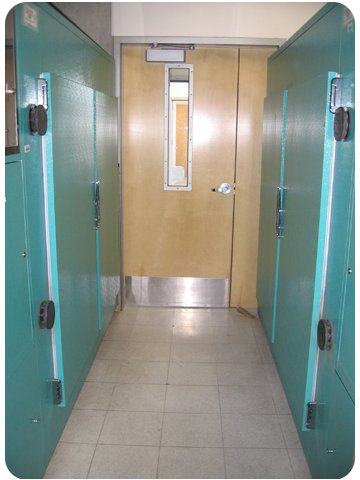
a) two Conviron plant growth chambers (first photo below),
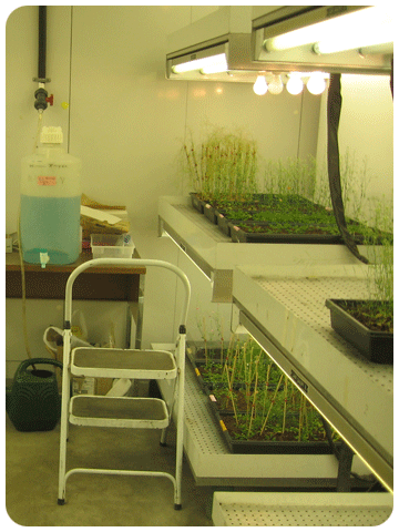
b) a walk-in plant growth room (with >100 sq ft of growth space; second photo below) equipped with LED Grow Lights and
c) >75sq ft of plant growth space in the University of Arizona greenhouses.
|
|
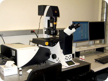
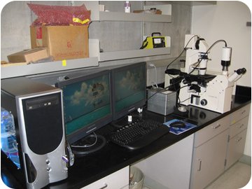
3. We perform time-lapse imaging using either a Leica SP5 Laser Scanning Confocal Microscope (photo above) or a Zeiss axiovert 100 fluroescent microscope (photo below). These microscopes are equipped with an automated X, Y, Z stages and shutter to generate multiple time-lapse movies using bright light plus green and red fluroescent laser lights. The microscopic observations are captured either by photo multiplier unit (in case of a confocal microscope) or by a Retiga CCD digital camera (in case of axiovert), stored on dedicated Image work stations (PC operated by Windows NIT with >100GB disk space) and analyzed using Leica, Metamorph and ImageJ image analyses softwares.
We set up multiple in vitro assays and then use the software to mark the position of each ovule (maximum capability = 96). Then, we utilize the software to capture a Z stack of images (covering up to 60 microns) of each marked ovule in red, green and bright field channels, once in every 10 minutes for 6-8 hours. The software drives all the stages to achieve these tasks and complete imaging 20 ovules in about 10 minutes, and return to resume this process all over again. At the end of the experiment, the time-lapse images for each ovule are assembled using Leica, Metamorph and ImageJ softwares.
4. Besides these, we have access to university-wide research facilities such as DNA sequencing, scanning and transmission electron microscopy, mass spectrometry and proteomics facilities. |
|


